×

Human Tumor Necrosis Factor α (TNF-α) ELISA Kit

Citations(2) Uniprot : P01375
  • Cat.No.: AE13959HU

  • Reactivity: Human (Homo sapiens)

To Purchase AE13959HU

Size:
48T 96T
Price: 查询

Reagent Preparation


Results demonstration


Assay Procedure Summary


Certificate


Product Details

种属 Human (Homo sapiens)
UniProt号 P01375
缩写 TNF-α
别名 TNF; DADB-70P7.1; DIF; TNF-alpha; TNFA; TNFSF2; APC1 protein|TNF superfamily; member 2|TNF; macrophage-derived|TNF; monocyte-derived|cachectin|tumor necrosis factor alpha; TNFSF7
线性范围 15.6-1000 pg/mL
灵敏度 10.4 pg/mL
样本类型 Serum, Plasma, Other biological fluids
检测方法 Sandwich
分析方法 Quantitive
反应时间 1-4.5h
样本体积 1-200 μL
检测波长 450 nm

原理

This assay employs a two-site sandwich ELISA to quantitate TNF-α in Human serum, plasma. An antibody specific for TNF-α has been pre-coated onto a microplate. Standards and samples are pipetted into the wells and any TNF-α present is bound by the immobilized antibody. After removing any unbound substances, a biotin-conjugated antibody specific for TNF-α is added to the wells. After washing, Streptavidin conjugated Horseradish Peroxidase (HRP) is added to the wells. Following a wash to remove any unbound avidin-enzyme reagent, a substrate solution is added to the wells and color develops in proportion to the amount of TNF-α bound in the initial step. The color development is stopped and the intensity of the color is measured.


产品概述

TNFa is synthesized as a 26 kDa, type II transmembrane protein that is 233 amino acids in length. It contains a 30 amino acid (aa) cytoplasmic domain, a 26 aa transmembrane segment, and a 177 aa extracellular region. TNFa is assembled intracellularly to form a transmembrane, non-covalently-linked homotrimeric protein. The 157 aa residue soluble form of TNFa (sTNF-α is released from the C-terminus of the transmembrane protein through the activity of TNFa-converting enzyme (TACE), a membrane-bound disintegrin metalloproteinase. Rat cells known to express TNF-αinclude B cells, colonic columnar epithelial cells, NK and CD3 CD56 hepatic natural T cells, macrophages, monocytes and monocyte-derived dendritic cells, CD4 and CD8 T cells, mast cells, neutrophils, keratinocytes, plasma cells, and adipocytes.


组份


Reagents

Quantity

Reagents

Quantity

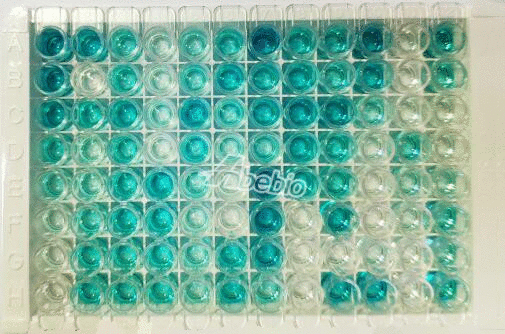
Assay plate (96 Wells)

1

Instruction manual

1

Standard (lyophilized)

2

Sample Diluent

1 x 20 mL

Biotin-Conjugate (concentrate 100 x)

1 x 120 μL

Biotin-Conjugate Diluent

1 x 12 mL

Streptavidin-HRP  (concentrate 100 x)

1 x 120 μL

Streptavidin-HRP Diluent   

1 x 12 mL

Wash Buffer (concentrate 25 x)

1 x 20 mL

Substrate Solution

1 x 10 mL

Stop Solution

1 x 6 mL

Adhesive Films

4


特异性

This assay has high sensitivity and excellent specificity for detection of Human TNF-α. No significant cross-reactivity or interference between Human TNF-α and analogues was observed.


回收率

Matrices listed below were spiked with certain level of recombinant Human TNF-α and the recovery rates were calculated by comparing the measured value to the expected amount of Human TNF-α in samples.

Sample Type

Number

Recovery range (%)

Average(%)

Serum

10

90-101

96

EDTA plasma

10

89-97

93

Heparin plasma

10

91-99

95


精密度

Intra-assay Precision (Precision within an assay) 
Three samples of known concentration were tested twenty times on one plate to assess intra-assay precision.
Inter-assay Precision (Precision between assays) 
Three samples of known concentration were tested in forty separate assays to assess inter-assay precision.
CV (%) = SD/meanX100
Intra-Assay: CV<8% 
Inter-Assay: CV<12%


线性

The linearity of the kit was assayed by testing samples spiked with appropriate concentration ofHuman TNF-α and their serial dilutions. The results were demonstrated by the percentage of calculated concentration to the expected.

Sample Type

1:2

1:4

1:8

1:16

Serum

78-89%

81-99%

92-103%

95-105%

EDTA plasma

91-101%

90-98%

93-101%

91-98%

Heparin plasma

92-103%

93-102%

92-99%

91-101%


稳定性

The stability of ELISA kit is determined by the loss rate of activity. The loss rate of this kit is less than 5% within the expiration date under appropriate storage condition. The loss rate was determined by accelerated thermal degradation test. Keep the kit at 37°C for 4 and 7 days, and compare O.D.values of the kit kept at 37°C with that of at recommended temperature. (referring from China Biological Products Standard, which was calculated by the Arrhenius equation. For ELISA kit, 4 days storage at 37°C can be considered as 6 months at 2 - 8°C, which means 7 days at 37°C equaling 12 months at 2 - 8°C).


样本收集及储存

Serum: Use a serum separator tube (SST) and allow samples to clot for two hours at room temperature or overnight at 2 - 8°C before centrifugation for 15 minutes at 1000 × g. Remove serum and assay immediately or aliquot and store samples at ≤ -20°C. Avoid repeated freeze-thaw cycles. Plasma: Collect plasma using EDTA, or heparin as an anticoagulant. Centrifuge for 15 minutes at 1000 × g at 2 - 8°C within 30 minutes of collection. Assay immediately or aliquot and store samples at ≤ -20°C. Avoid repeated freeze-thaw cycles. Other biological fluids: Centrifuge samples for 20 minutes at 1000 × g. Remove particulates and assay immediately or store samples in aliquot at -20°C or -80°C. Avoid repeated freeze/thaw cycles.


注意事项

Store at 2-8°C. Please refer to Instruction Manual.
 
  1. ZEB2 knockdown inhibits interleukin-1β-induced cartilage degradation and inflammatory response through the Wnt/β-catenin pathway in human chondrocytes IF: 2.100
    Osteoarthritis (OA) is a degenerative disease of the joints characterized by inflammation and cartilage degeneration.
  2. Roles of TREM2 in degeneration of human nucleus pulposus cells via NF‐κB p65 IF: 2.959
    Nuclear factor (NF)-κB p65 plays a key role in the development of intervertebral disc degeneration (IDD). Herein, we found that messenger RNA levels of human triggering receptor expressed on myeloid cells‐2 (TREM2) and NF‐κB p65 were upregulated and strongly positive correlated (r2 = 0.299, P = 0.0126) in nucleus pulposus (NP) tissues of patients with IDD.